Computed Tomography applications for museum research
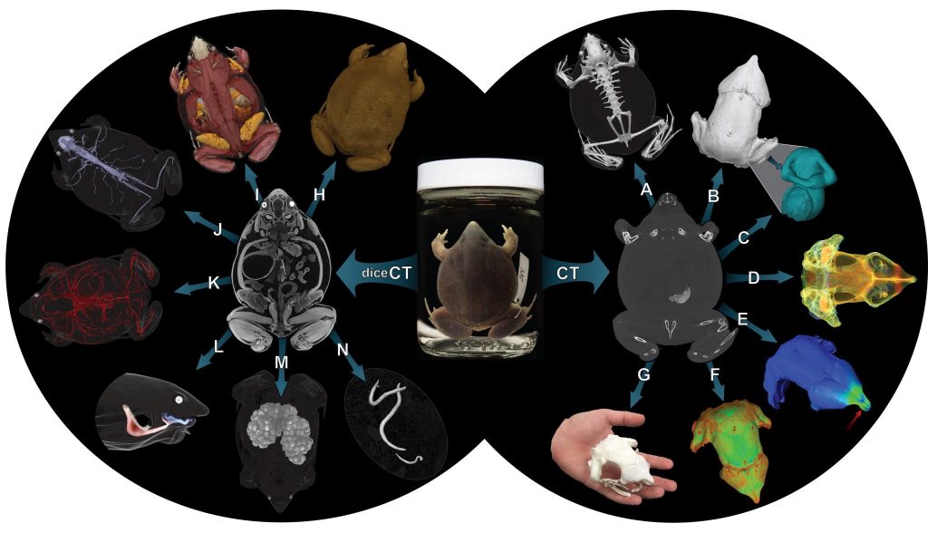
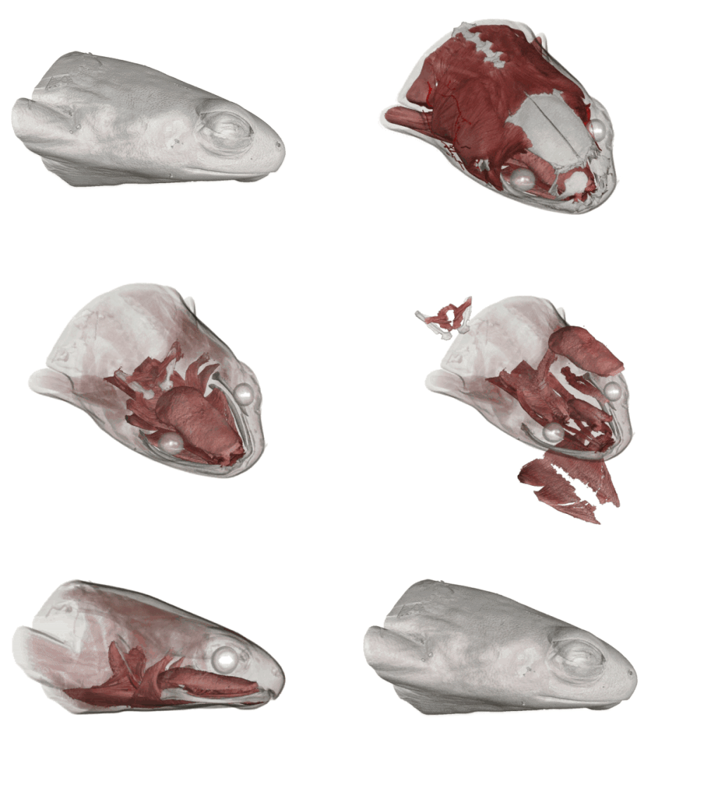
High Resolution Computed Tomography, also known as X-ray microtomography/ nanotomography, is a nondestructive imaging technique that allows the ultrahigh-fidelity reconstruction and visualization of external and internal features of non-living objects. This technique has the potential to revolutionize collections and collections-based research, and has many applications in an ever-growing range of biological systems. Although HRCT has traditionally been limited to visualizing denser material (bone, fossil etc.), improved hardware and new contrast-enhancing techniques now allow this technology to visualize many different types of soft anatomy in exquisite detail.
The University of Florida Nanoscale Research Facility operates a Phoenix V|Tome|X M dual-tube nano-CT system and a ZEISS Versa 620 X-ray Microscope. The Museum uses these systems extensively for research, and has scanned everything from tiny bird lice insects to fossil alligator skulls. If you would like to receive training in the use of these machines, or are interested in having the NRF staff scan your samples, you can begin both processes here.
Processing CT data at the 3D lab
The datasets produced by the UF CT systems often require extensive post-processing. The 3D Lab contains four high-end segmentation computers with a range of reconstruction and 3D editing software, including VGStudioMax 3.3, a powerful voxel-analysis software package from Volume Graphics. Please check availability of the 3D lab resources here, and contact Edward Stanley to arrange time on the schedule. We offer introductory, intermediate and advanced CT segmentation workshops each semester, as well as graduate (ZOO6927) and undergraduate (ZOO4926) “CT for Biologists” classes, which are offered every year in the fall.
Recovering CT data from UF specimens
The Florida Museum of Natural History is the lead institution of the oVert Thematic Collections network, a multi-institution, NSF-funded initiative that is digitizing and sharing CT data from every vertebrate genus available in U.S. collections. The resulting tomogram stacks and select shape files are freely available on Morphosource.

