Lesson: Sawfish Skin: Adaptation for Life Underwater
Lesson Summary:
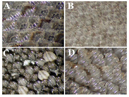
This is a teaching activity to show students shark skin in detail under a dissecting microscope. They may be able to see some differences in skin structure and appearance of skin samples from different species of sharks. (Sawfish skin samples are not available due to the endangered status of these fish, shark skin is very similar in form and function to sawfish skin.)
Vocabulary:
Dermal denticles, hydrodynamic, predators, dissecting scope

Background Information:
Shark skin is very similar to sawfish skin. It is made of many hard, tooth-like structures called dermal denticles. The denticles are pointed and grooved and have different shapes depending upon the species of shark and location on the shark’s body. As the shark grows, the denticles do not increase in size, but rather the shark grows more denticles. The denticles help the shark swim more quickly due to the hydrodynamic shape that allows the water to flow more freely over the surface of the shark. The roughness of the skin also helps protect the shark from potential predators.
The skin is covered with dermal denticles. Notice in the photo that the denticles are arranged in the same direction. This enables the shark to swim more quickly through the water than it could otherwise. This is also why when you stroke the skin from head to tail it feels very smooth but like sandpaper if stroked the opposite direction.
Materials:
Teacher’s activity guide
Shark skin samples
Dissecting microscopes
Paper to make species labels
Procedure:
First, set up dissecting microscopes. Then break the class into groups and give each group a shark skin sample. Next have each group place the skin sample under the scope, with each group making a label for the shark species next to the microscope. Then have each group move from scope to scope to see the differences in the denticles between the three species.
Discussion Questions:
- What purpose do dermal denticles serve on the skin of elasmobranches (sharks, rays, skates, and sawfish)?
- When you touch the skin sample, how does it feel? Try stroking the skin in the opposite direction, does it feel the same?
- Can the students guess what direction the head of the shark would be and where the tail would be and why?
- Why are we using shark skin instead of sawfish skin?