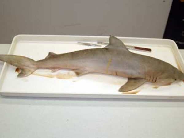
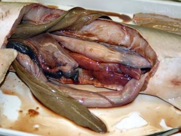
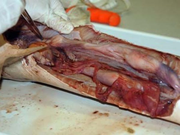
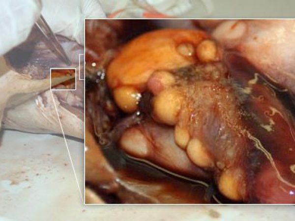
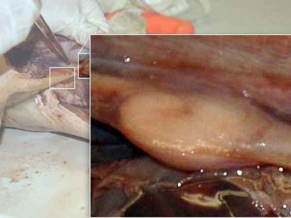
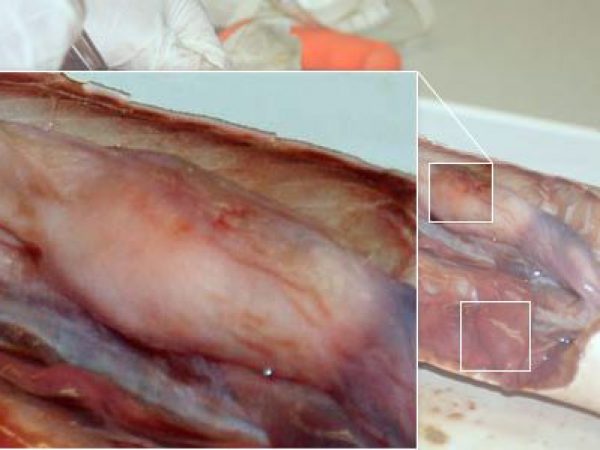
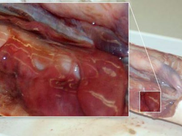
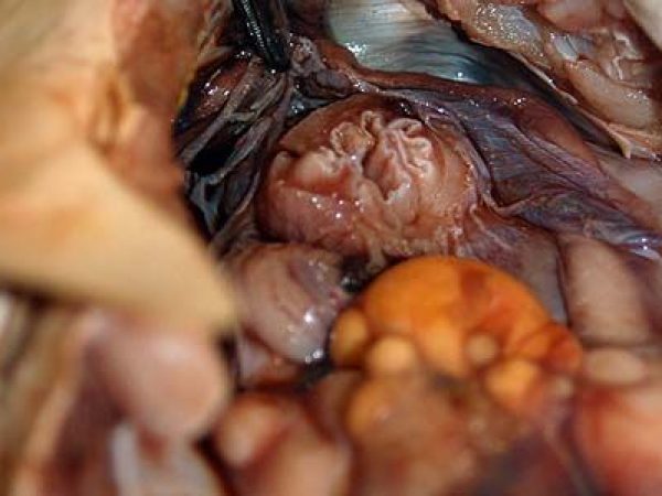
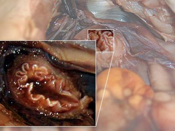
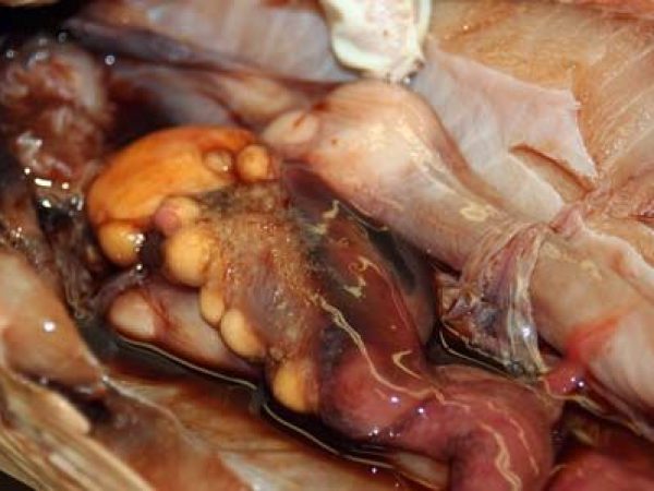
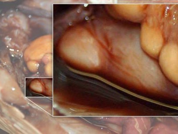
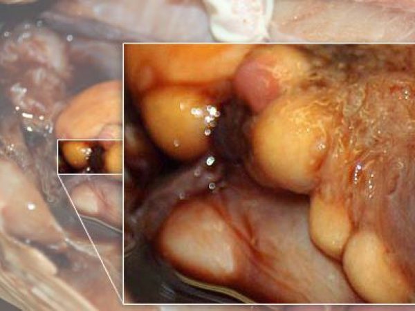
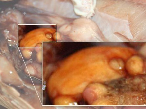
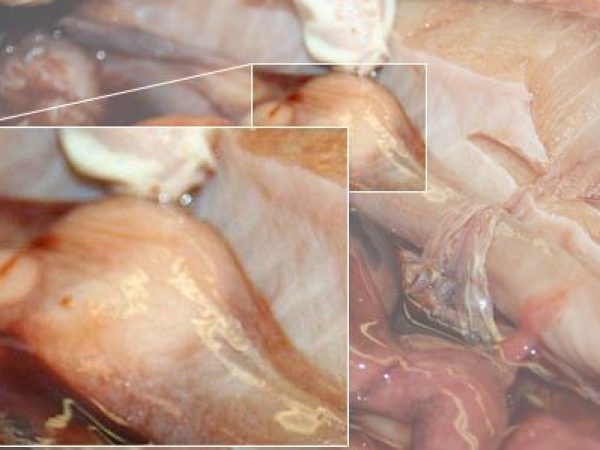
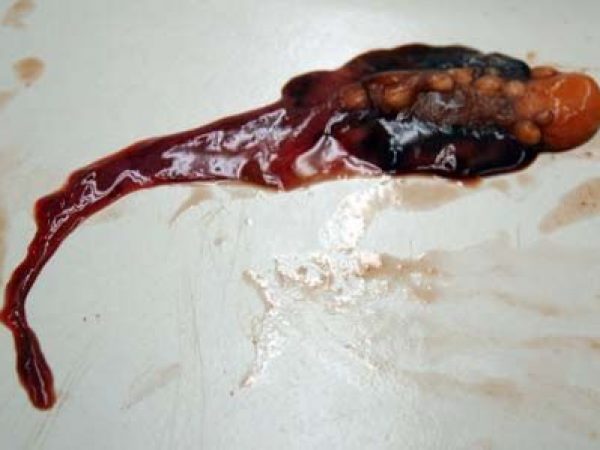
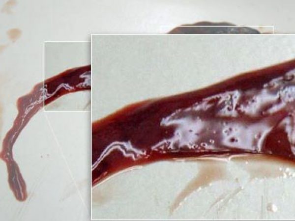
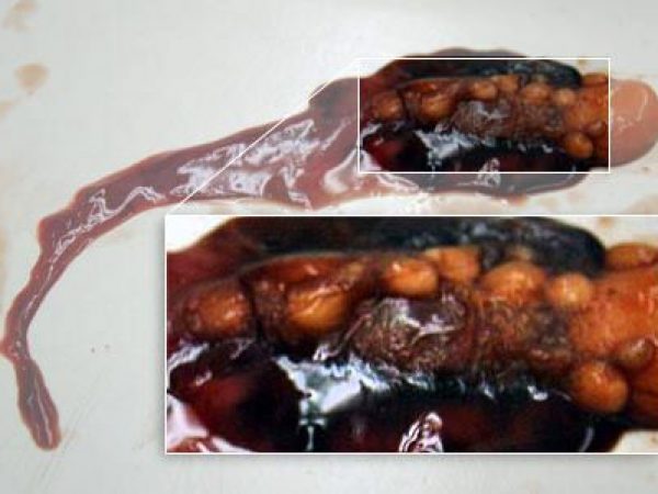
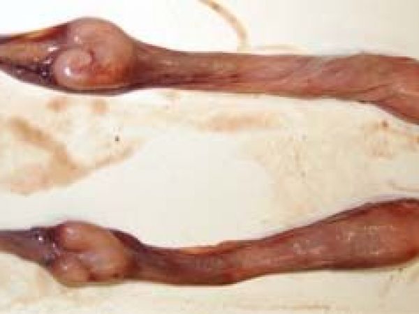
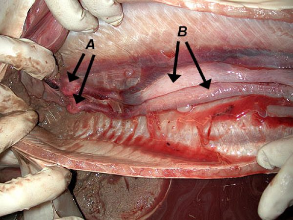
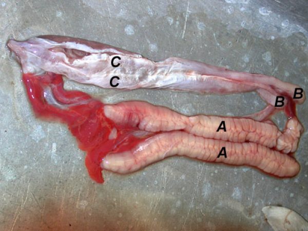
This gallery of images documents a dissection of a small, female blacknose shark (carcharhinus acronotus), focusing on the reproductive organs.
–Click for larger images–
 Female blacknose shark (carcharhinus acronotus)
 Internal view of the body cavity with digestive organs still present.
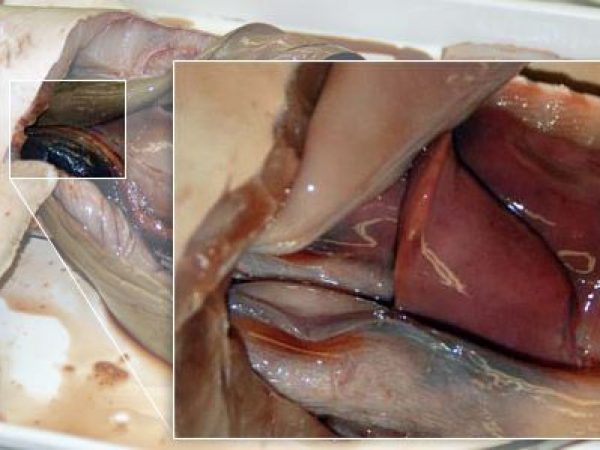
 Close up: common urogenital duct.
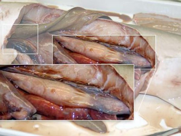
 Close up: oviduct.
 Internal view of the body cavity with the major digestive organs removed.
 Close up: ovary.
 Close up: nidamental gland.
 Close up: uterus.
 Close up: epigonal tissue.
 Internal view showing the ovary in relation to the esophagus.
 Close up: esophagus.
 Internal view focusing on the ovary, eggs, and right and left nidamental glands.
 Close up: right nidamental gland.
 Close up: eggs.
 Close up: ovary.
 Close up: left nidamental gland.
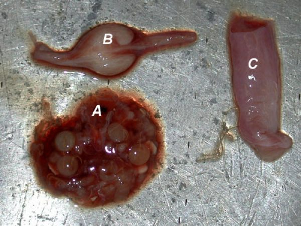
 Removed ovary, eggs, and epigonal tissue.
 Close up: removed epigonal tissue.
 Close up: removed eggs.
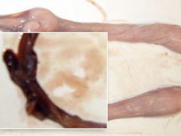
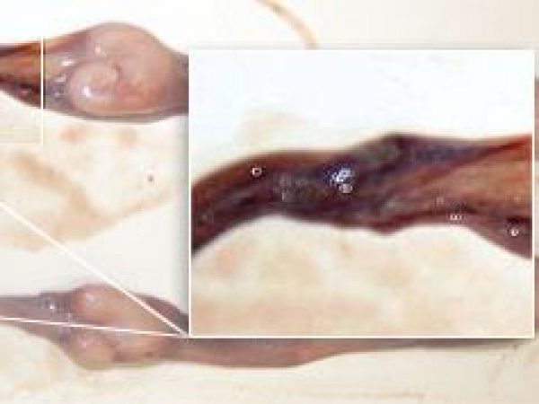
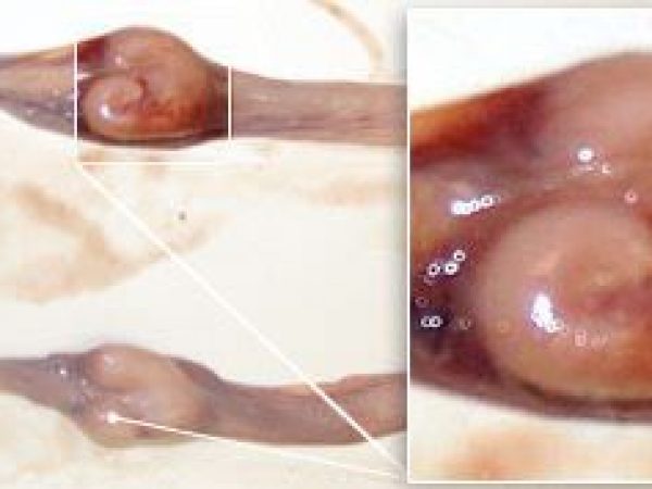
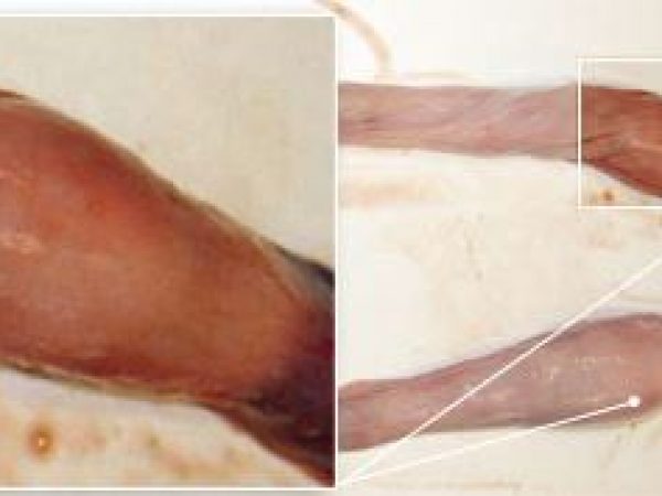
 Removed reproductive tract, highlighting the ostium and pairs of oviducts, nidamental glands, and uteri.
 Close up: ostium.
 Close up: right and left oviducts.
 Close up: right and left nidamental glands.
 Close up: right and left uteri.
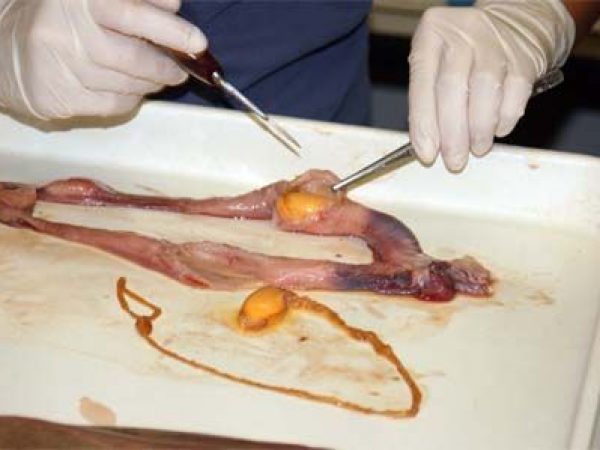
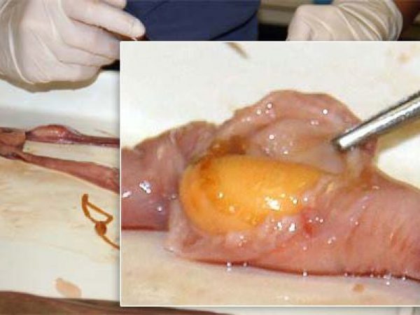
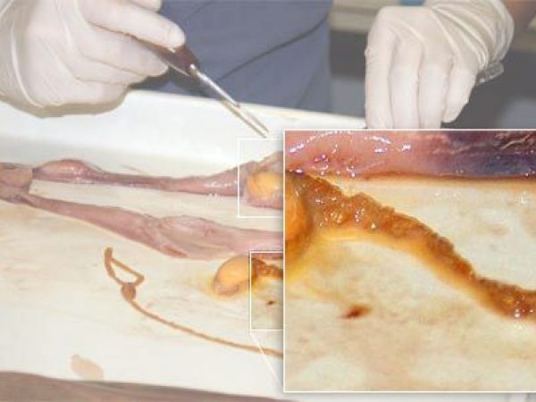
 Dissector removing a fertilized egg from the uterus.
 Close up: fertilized egg.
 Close up: egg membrane.
Female Shark Reproductive Images
Male Shark Reproductive Images
