Some of the digital images produced from the University of Florida Herbarium (FLAS) specimens were prepared using several types of scanners and digital cameras which are no longer in use. These are summarized below.
The earliest efforts were supported by the University of Florida Libraries Digital Library Center and the Florida Center for Library Automation (FCLA). We are dearly indebted to the personnel at the Digital Library Center and FCLA for those ground-breaking efforts. Project listings prioritized selected categories of plants. These included: Introduced Florida Plant Species, Potentially Poisonous Florida Plant Species, FLAS Type Specimens, Insectivorous Florida Plant Species, John Bartram’s Botany: St. Augustine to Picolata and Floristic Inventory of Kanapaha Botanical Gardens project specimens.
Image file names were based on the accession number of the herbarium specimen. Image files were served via the Zoomify® viewer utilizing XML and Java script, or an alternate Zoomify® Adobe Flash viewer. From ca. 2010–2023, images were prepared with a TTI-Repro-Graphic Workstation 3040 with a Digiflex 67ei Sinar 75H camera back and a Sinar Evolution 75H Multi Shots Digital Back System (33MP). These images are distinguished by the ruler and color charts present on the left side of the sheets. We estimate the image resolution to be ca. 440 ppi. Detailed specifications are as follows:
Sinar Evolution System with Sinar CaptureFlow Software and TTI-Repro Graphic Workstation (discontinued)
Camera: Sinar Evolution 75H multi shots 33MP digital back.
Lighting-table-camera mount: TTI-Repro-Graphic Workstation 3040 and Digiflex 67ei.
Capture Software: Sinar CaptureShop.
Image Resolution: ca. 400 ppi / Acquistion Throughput: around 1 minute per specimen.
Workflow and Use Instructions: PDF Required a Mac cpu with an OS around ca. 2010 (OS 10.6.4).
Cost: See this study
The following video, produced by iDigBio, features the Sinar System:
Herbarium digitization video by iDigBio
- Workstation: 25″x32″x24″ High, worktable. 60″ Column height; Motorized Camera elevation with variable speed remote control, lead screw driven; Digital counter and camera mount. Camera: TTI Digiflex 67ei Modified to incorporate Sinar CMV Lenses with electronic shutter for live preview and media positioning. Precision hi resolution lead screw focusing. Interface mounting plate for the 75H digital camera back. Lighting: Quartz halogen TTI DL-400 day light color temperature 5500 Kelvin (8) lamp system, 4 lamps per side with integral dichroic reflector and forced air cooling; TTI new lighting mounting system, which telescope IN/OUT, UP/DOWN and Angular Pivoting, all with Engraved Markings for repeatable set-ups and additional flexibility. TTI will supply a shadowless diffusion system for intricate specimens lighting. Lights turn On/Off via footswitch. Voltage Stabilizer supplied by TTI will maintain light color temperature & light level consistent during captures. Foot Print: 89″W X 35″D X 96″H; Electrical: Dedicated line 120VAC 50/60Hz-20 Amps Circuit Breaker.
- Camera: Sinar Evolution 75H Multi Shots Digital Back System including 33MP Digital Back, 95MB RGB 24 BIT TIFF,190MB RGB 48 BIT TIFF file in one or four shots mode. Files can be scaled up through Sinar Capture Shop software to 244MB RGB 24 BIT TIFF and to 488MB RGB 48 BIT without loss of details. Live video and focus, 90mm Sinaron Digital CMV Lens.
The earliest images on our site (circa 2000-2006) were prepared by the staff of the George A. Smathers Libraries Digital Library Center. Despite being created in the early 2000’s, these images are among the highest resolution on our site. They also took an average of 20 minutes each to acquire with the “top-of-the-line” hardware. They may be recognized by the presence of a small white 5 cm. ruler and no color chart. They were acquired using a ZBE Satellite large format stationary camera equipped with a PowerPhase ARI cameraback, 135mm Rodenstock lens and daylight filter. A Macintosh (PowerMac G4) computer system with 17 gigabytes of storage and ~ 800 megabytes of active memory was used for processing. The camera images were matched with a 1.8 gamma monitor, relative colorimetrics and the “BEST” quality for rendering profiles. Colorimetrics were calibrated using a Kodak Q-60R1 target per ANSI IT8.7/2-1993(1999:04). Intermediate processing was achieved with Adobe Photoshop 5.0. The final archives were recorded on CD’s with accessible images made available on the Internet via the Lizardtech viewing source Mr. SID. from the Florida Center for Library Automation web server. All of those original images have subsequently been moved to the FLMNH server. Here are specifications as provided by the DLC:
- Every effort has been made to preserve images as accurately as the original. Specimens are photographed at a 1:1 ratio, meaning camera height, DPI (ppi), and output image are all consistent with the original. This means that an 11.5 x 17.5 in. specimen sheet is actually preserved as an 11.5 x 17.5 in. final image. A measurement scale has been included in the images as a reference. In most cases post-production manipulation is unnecessary, but occasionally (usually those with several centimeters of depth of field) it is necessary to use the sharpening and brightness/contrast filters in Adobe Photoshop 5.0.
- Digital files are acquired using Phase-One camera software with a gray balanced standard film curve and ISO sensitivity of 400. Specimens are initially recorded as Macintosh (3 vector) images with final format being TIFF files with color depth at 24/16 millions of colors. Generally entire sheet specimens can take anywhere from 5 to 10 minutes (in some cases up to 25 min.) to acquire, have an average of 450 DPI (ppi), and file sizes around 140 megabytes. Close-up’s can take anywhere from 45 seconds to 5 minutes and may exceed 3500 DPI (ppi), and can range from 500 bytes to 80 megabytes in size.
Staff and volunteers in the University of Florida Herbarium also acquired images of selected specimens on a flatbed scanner, Microtek Scanmaker 9600XL. Those specimens were scanned at 400 ppi and saved in tif format (.tif). These images may be recognized by the presence of a small white 6 in./15 cm. ruler and no color chart. Most of the specimens available in the Floristic Inventory of Kanapaha Botanical Gardens project are prepared by this method.
Digital images of plant materials taken with consumer model digital cameras prior to pressing are being added to compliment the specimens. Most of these images are prepared and posted in jpeg (.jpg) format.
HerbScan / Epson Scanner System
This system is an Epson Scanner mounted upside down. This equipment was used to scan high resolution images for JSTOR Global Plants (plants.jstor.org). It mostly serves as a backup imaging system now, or is sometimes used for other imaging needs.
Equipment: Epson Expression 10000 XL flatbed scanner with HerbScan stand.
Capture Software: Epson Scan with post processing in Adobe Photoshop.
Image Resolution: 600 ppi (1200 ppi) / Acquistion Throughput: around 3 minutes per specimen.
Workflow and Use Instructions: PDF
Microtek ScanMaker 9600XL/9800XL
In the early 2000s this scanner was used to scan some specimen images.
Nikon System
- Camera: Nikon D810 36MP digital SLR camera (body).
- AC Power Adapters for the Nikon D810: Nikon EH-5B AC Adapter (requires EP-5B); Nikon EP-5B Power Supply Connector.
- Camera and Imaging Software: Nikon Camera Control 2; Adobe Creative Suite (Lightroom and Photoshop); Adobe DNG Converter.
- Camera Lens: Sigma 50 2.8 EX DG MAC F/Nikon AF-D USA. This lens works with the Photo e-Box and has a field of view that closely matches the herbarium specimen.
- Miscellaneous supplies: microfiber cloth for lens cleaning, gaffer tape, Jobu Design dual axis bubble level.
- Copy Stand: We are using our existing Bencher Copymate II stand. We removed the daylight florescent lights and stored them because we are using the Photo e-Box with LED lights (below). The iDigBio guide lists other copy stand options.
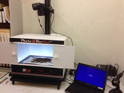 Photo e-Box: model 1419, NYBG modified, Photo-e-Box from OR Technologies.
- Barcodes: The UF Herbarium uses custom barcodes printed by Computype, Inc.: Digitek DC1551 (P236 – Adhesive, L107 .5 mil clear matte laminate, B274 polypropylene base material). Other barcode vendors include Watson Label Products.
- Barcode Reader: needed to read barcodes which are used for file naming when imaging.
- The Photo e-Box has a white base. We prefer a black magnetic base so that we can see the edge of the sheets and use magnets to secure uneven specimens. One of our volunteers, Steve Stevens, covered a magnetic board and rulers with black vinyl. These were cut to fit the box so we have two edges to align the specimens against.
- The Photo e-Box tends to slide on the copy stand. Our volunteer made a board with raised edges and a not skip bottom to place the Photo e-Box on.
- White magnets are used to flatten loose or bent areas of sheets. We tried white magnets purchased from K&J Magnetics. Unfortunately, they are neodymium magnets and much too strong and hazardous to use. Regular magnetic disks that are 1/2″ or 3/4″ in diameter may covered with white stickers and used.
- Magnetic Rulers are used along the edge of the specimen image. We purchase 6″/15cm black magnetic rulers, A-6206M from Arrowhead Forensics. 2″/5cm rulers are also available.
- Color calibration/separation and white balance guide. There are a wide variety available:
Tiffen Color Separation Guide with Grey Scale, 8″ Size, #Q-13: useful to use as a border along the side of herbarium sheets.
Digital Transitions ColorGauge Nano Target: this guide is postage stamp size (9/16″ x 13/16″) and very durable. It is expensive (ca. $200), but given how easy it is to fit in an image, we have settled on using it as have quite a few other herbaria.
X-Rite Mini ColorChecker Card P/N 50111: 2 1/4″ X 3 1/4″: useful to place in bare areas on herbarium sheets. This was the standard guide used by most institutions for the Global Plants Initiative project. This guide may have been discontinued? It doesn’t seem to be available. Perhaps, the CameraTrax 24 ColorCard 2X3 with 12% white balance will work ok?
Configuration of the camera and software is detailed in the Camera Configuration page.
Useful Resources
Camera Configuration (via Nikon Camera Control Pro 2 Software)
Camera: Nikon D810 with Sigma 50 2.8 EX DG MAC F/Nikon AF-D USA lens.
Positioning the Camera and Photo e-Box
The bottom of the light stand camera holder is set at “82”. This places the camera lens ca. 25 1/4″ from the light box platform. This camera height and Photo e-Box position is situated so that the herbarium sheet, ruler and color chart fill the field of view with as little blank space as possible.
Nikon Camera Control Pro 2 Software
Out-of-the-box, the Camera Control 2 software did not support the Nikon D810. We had to get the latest version from the Nikon web site.
Useful Resource
Nikon Camera Control Pro 2 screens and settings
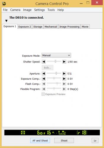
- Exposure: Manual
- Shutter: 1/60 sec
- Aperture: f/11
- Exposure compensation: 0 EV
- Flash compensation: 0 EV
- Flexible program: 0 steps
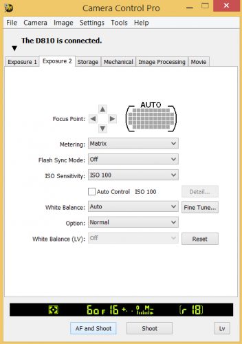 Exposure 2
- Focus point: Auto
- Metering: Matrix
- Flash sync mode: Off
- ISO sensitivity: ISO 100
- White balance: Auto (fine tune)
- Option: Normal
- White balance (LV): Off
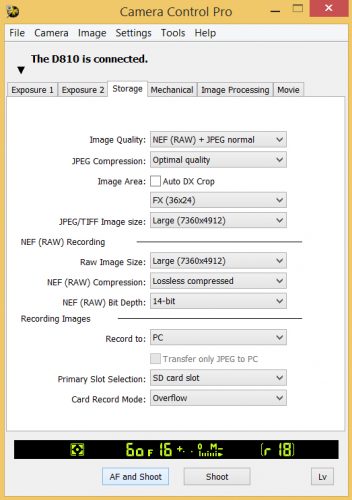
- Image Quality: NEF (RAW) + JPEG normal
- JPEG Compression:
- Image Area: Auto DX Crop (unchecked), FX (36×24)
- JPEG/TIFF Image Size: Large (7360×4912)
- NEF (RAW) Recording
- Raw Image Size: Large (7360×4912)
- NEF (RAW) Compression: Losless Compressed
- NEF (RAW) Bit Depth: 14-bit
- Recording Images
- Record to: PC (transfer only JPEG to PC (unchecked)
- Primary Slot Selection: SD card slot
- Card Record Mode: Overflow
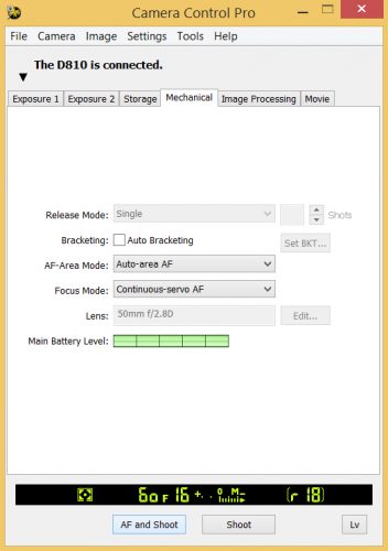
- Release Mode: Single
- Bracketing: Auto Bracketing (unchecked)
- AF-Area Mode: Auto-area AF
- Focus Mode: Continuous-servo AF
- Lens: 50mm f/2.8D
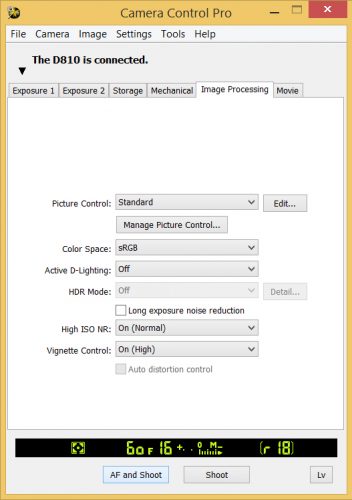
Picture Control: Standard
Color Space: sRGB
Active D-Lighting: Off
HDR Mode: Off
Long exposure noise reduction: Off
High ISO NR: On (Normal)
Vignette Control: On (High)
Auto distortion control Unchecked
Image File Naming Conventions (historic)
FLAS Accession Number + Image Designation + photo number
E.g.: P555a1, 112555g1, 112555g2
FLAS Accesssion Number – an optional capital letter, from 1 to 6 numbers, an optional capital letter. E.g., 112555, 3, P444, B0444, 175995A
Image Designations – a single lower case letter based as follows:
| Letter | Image of |
|---|---|
| a | sheet (entire specimen) |
| b | fragment packet contents |
| c | plant close-up (formerly, bottom of plant, may include rootstock). This may be leaves, roots or sections of plant. |
| d | – [top of plant (may include inflorescence / infructescence): deprecated, use c] |
| e | – [leaves: deprecated, use c] |
| f | – [leaf upper side: deprecated, use c] |
| g | sheet prepared for Global Plant Initiative, plants.jstor.org [leaf lower side: deprecated, use c] |
| h | – [stem: deprecated, use c] |
| i | inflorescence |
| j | flower |
| k | infructescence |
| m | fruit |
| n | seed (if large) |
| p | pollen or spore(s) |
| s | sheet for SERNEC project with Photo e-box |
| v | seed vial contents |
| w | – [plant close-up (e.g., if small plants or region of sheet), deprecated, use c] |
| x | photo of fresh plant or plant in the field, typically, field photos by collectors names by collector image tag and collection number |
| z | photo on sheet or of textual material accompanying the specimen |
Number – the image number for an image designation category for the sheet starting with one.
Credits for Initial Imaging Phases
Project and Web Page Design, Direction and Oversight
Priscilla Caplan, Florida Center for Library Automation
Stephanie Haas, Digital Library Center, University of Florida Libraries
Erich Kesse, Digital Library Center, University of Florida Libraries
Kent Perkins, University of Florida Herbarium, Florida Museum of Natural History
Digital Imagery of Specimens
Michael Bond, Digital Library Center, University of Florida Libraries
Erich Kesse, Digital Library Center, University of Florida Libraries
Herbarium Type Specimen Database
Kent Perkins, University of Florida Herbarium, Florida Museum of Natural History
Wendy Zomlefer, University of Florida Herbarium, Florida Museum of Natural History
–now, Curator, University of Georgia Herbarium
Computer Technical Support and Server Maintenance
Nicholas Bazin, Florida Museum of Natural History
Sarah Fazenbaker, Florida Museum of Natural History
Chris Cuevas, Florida Center for Library Automation
William Paine, Florida Museum of Natural History
Web Page Text
Kent Perkins, University of Florida Herbarium, Florida Museum of Natural History
Stephanie Hass, Digital Library Center, University of Florida Libraries
Priscilla Caplan, Florida Center for Library Automation
Erich Kesse, Digital Library Center, University of Florida Libraries
Michael Bond, Digital Library Center, University of Florida Libraries (Digital Imaging section)
Astrid Terman, Florida Center for Library Automation (Help section)
