GAINESVILLE, Fla. — Visitors to the Florida Museum of Natural History’s latest gallery exhibit can take a uniquely artistic look into the museum’s fish collection.

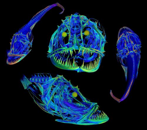
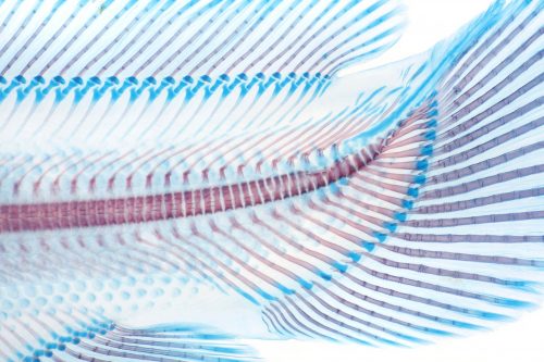
“Inner Beauty: Skeletons Revealed from the Museum’s Fish Collection” reveals rarely-seen views of the museum’s fish specimens. Vibrant, colorful images showcase the results of two techniques used in modern preservation science: clearing and staining and CT scanning.
“People can take a look at some of these really cool techniques and processes that usually happen behind the scenes and they never get to see,” said Zach Randall, manager of the Florida Museum’s imaging lab and creator of most of the images featured in the exhibit. “Science can be very intimidating to some people. The idea here is to explain the mystery behind some of these processes by using images and videos that people can connect with visually.”
The exhibit features a video of Randall demonstrating both clearing and staining and CT scanning, giving visitors a look at the process of these two skeletal preparation techniques. The actual specimens depicted in the scans are also on display, along with the pros and cons of both preservation methods. iPads with interactive skeleton models of a lined seahorse and shovelnose sturgeon give guests another uncommon look at life under the sea.

“When I saw Zach’s beautiful images, I was enthralled by them, but also fascinated by how the science was changing behind preservation and wanted to share that with our visitors,” said Florida Museum exhibit developer Tina Choe. “With advanced technology, museum specimens and objects are more accessible than ever for research, education, art and community outreach.”
Some of the images in this exhibit were created from CT scan data made available through the Florida Museum’s openVertebrate Thematic Collection Network (oVert), a collaborative initiative funded by the National Science Foundation. oVert provides thousands of open-access CT scans of specimens from museum collections across the country to the general public. The exhibit also highlights ways educators and artists have used these scans, such as 3D-printing snake and frog skulls for classrooms and creating a giant frog sculpture featured in a Carnegie Museum of Art exhibit.
For more information, visit www.floridamuseum.ufl.edu/exhibits/inner-beauty.
-30-
Writer: Nikhil Srinivasan, nsrinivasan@floridamuseum.ufl.edu
Sources: Tina Choe, tchoe@floridamuseum.ufl.edu; Zach Randall, zrandall@flmnh.ufl.edu
Media Contact: Kaitlin Gardiner, kgardiner@floridamuseum.ufl.edu